Research Grants


  • NIH Outstanding Investigator Award (R35), MIRA-ESI
  • Issued by the National Institutes of Health in 2022, Dr. Moradi has been awarded the NIH Outstanding Investigator Award for work on Physics-based characterization of functionally relevant protein conformational dynamics (R35GM147423) until 2027.
  • NSF I-Corps Award
  • Issued by the National Science Foundation, the I-CORPS award was given in 2021-2022 for a Physics-Based Binding Affinity Estimator (NSF IIP 2138667).
  • NSF CAREER Award
  • Dr. Moradi has been awarded the National Science Foundation CAREER Award in 2020 for work on Riemannian Reformulation of Collective Varaible Based Free Energy Calculation Methods (NSF CHE 1945465) until 2025.
  • NIH R01 Grant
  • Granted by the National Institutes of Health in 2024, Dr. Moradi is a co-I for work on Probing Real-time Conformational Dynamics and Allosteric Cooperativity of the HIV-1 Envelope Glycoprotein During Virus Entry in collaboration with Dr. Maolin Lu at The University of Texas at Tyler until 2028 (NIH R01-AI181600-01).
  • DOE Grant
  • Granted by the Department of Energy in 2020, Dr. Moradi was a co-PI for work on Protein Targeting to the Chloroplast Thylakoid Membrane: Structure and Function of a Targeting Complex" in collaboration with Dr. Colin Heyes at the University of Arkansas until 2023 (DE-FG02-01ER15161).
  • NIH R15 Grant
  • Granted by the National Institutes of Health in 2020, the lab recieved the NIH R15 Grant until 2023 for work on Molecular Characterization of the Influenza Hemagglutinin Mediated Membrane Fusion (NIH R15-GM139140-01).
  • NSF HDR Grant
  • Granted by the National Science Foundation in 2019, the lab recieved the NSF HDR Grant until 2022 for Collaborative Research: Atomic Level Structural Dynamics in Catalysts (NSF OAC 1940188).

    Cholesterol Dependence on the Conformational Changes of Metabotropic Glutamate Receptor 1 (mGluR1)

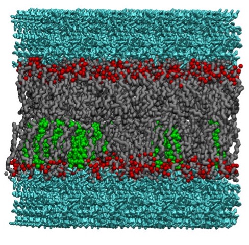
    MD Simulation of mGluR1 in Various Cholesterol Concentrations


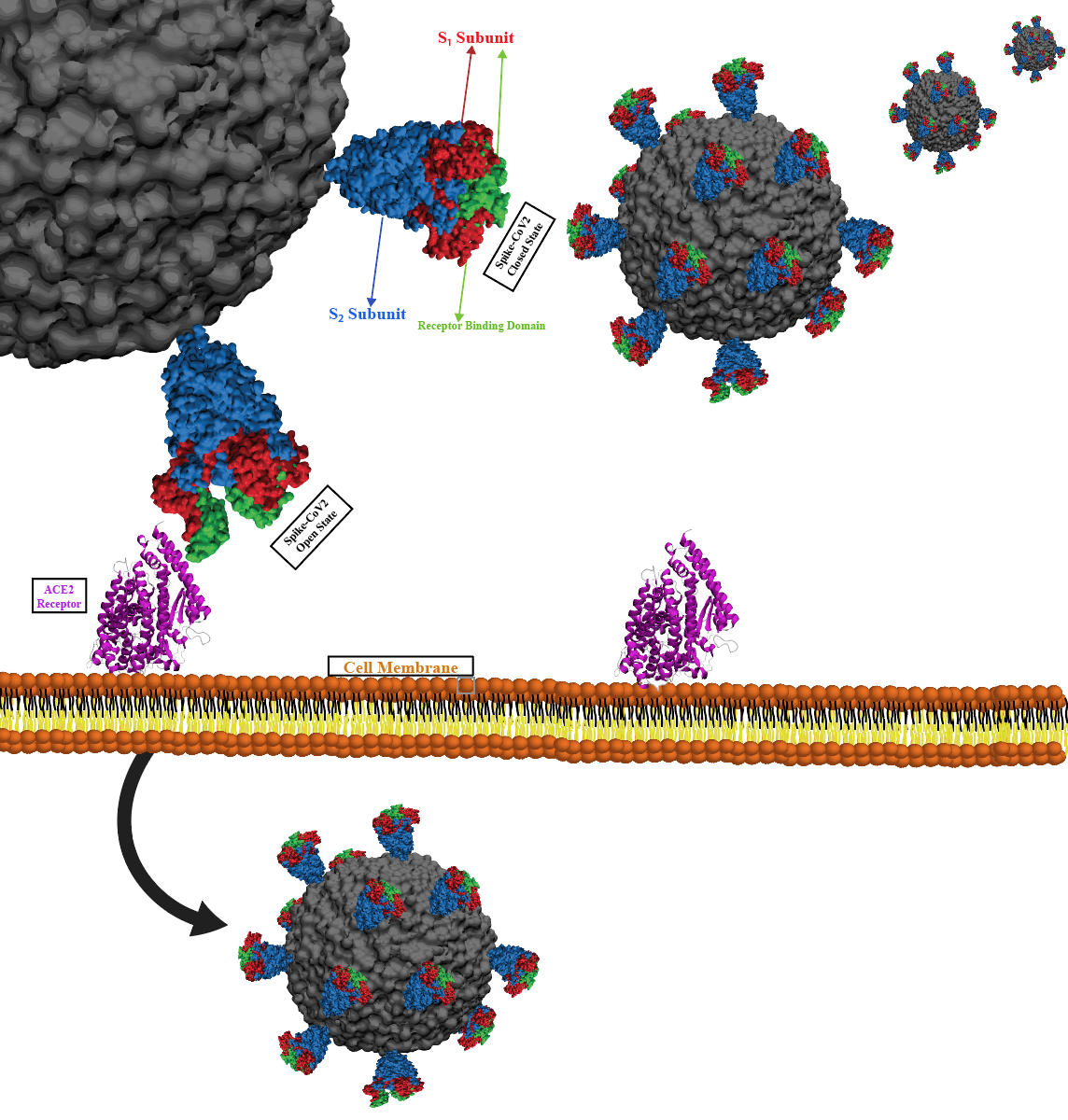
    GPCRs are extremely successful drug targets for treatment of diseases in medicine, with approximately 35% of approved drugs in the pharmaceutical market 
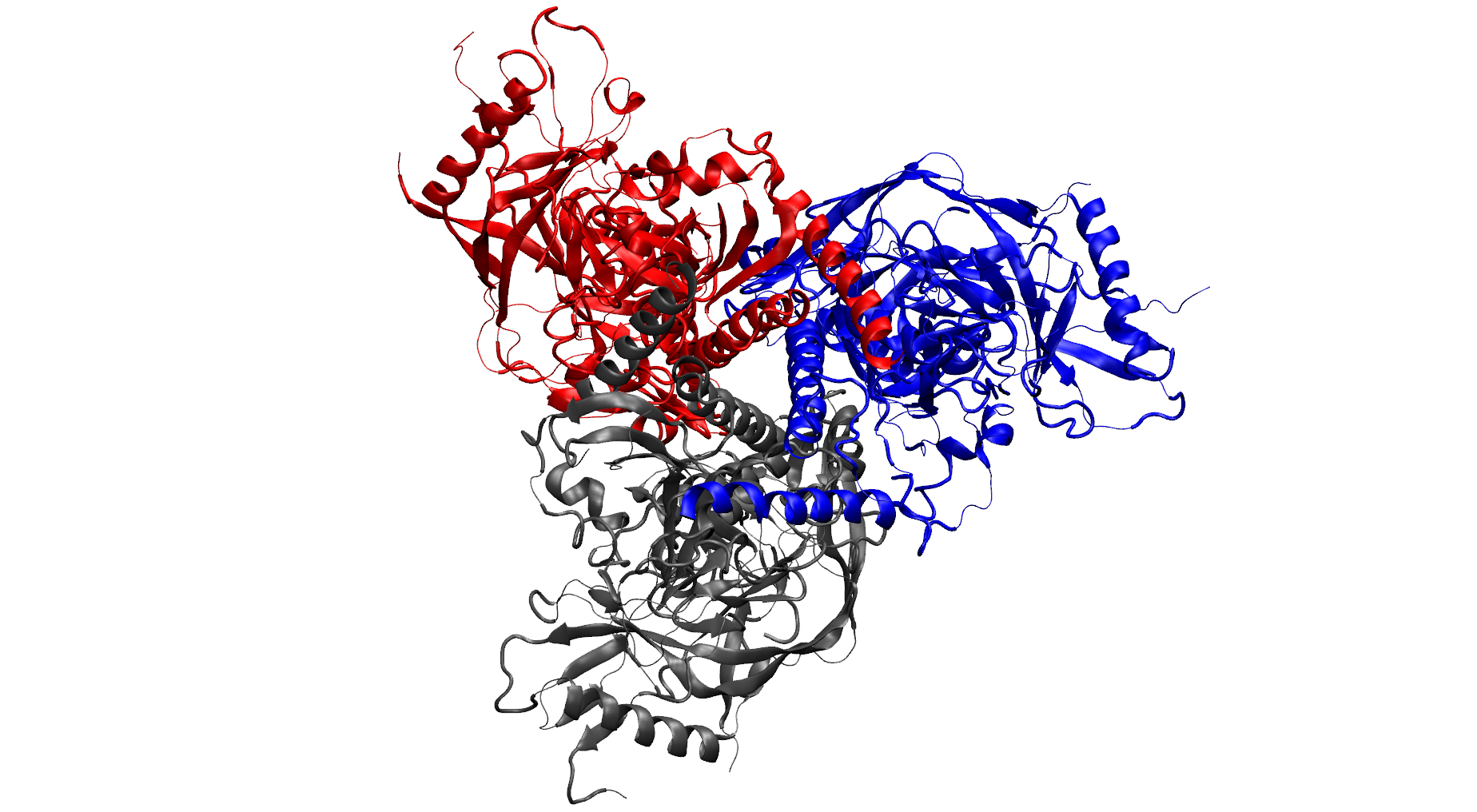
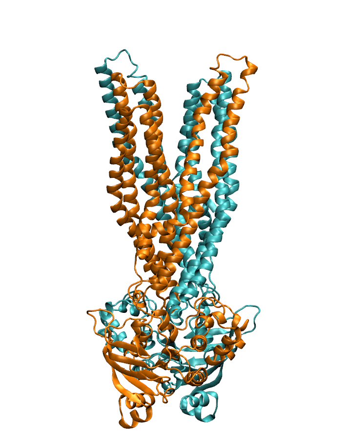
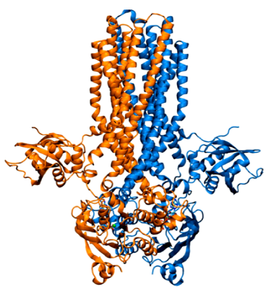
targeting GPCRs. Metabotropic glutamate receptors (mGluRs) are class C G-protein-coupled receptors (GPCRs) which exist as constitutive dimers. They play significant roles in regulating neurotransmission and activating excitatory synapses in the central nervous system. Besides the common GPCR-defining 
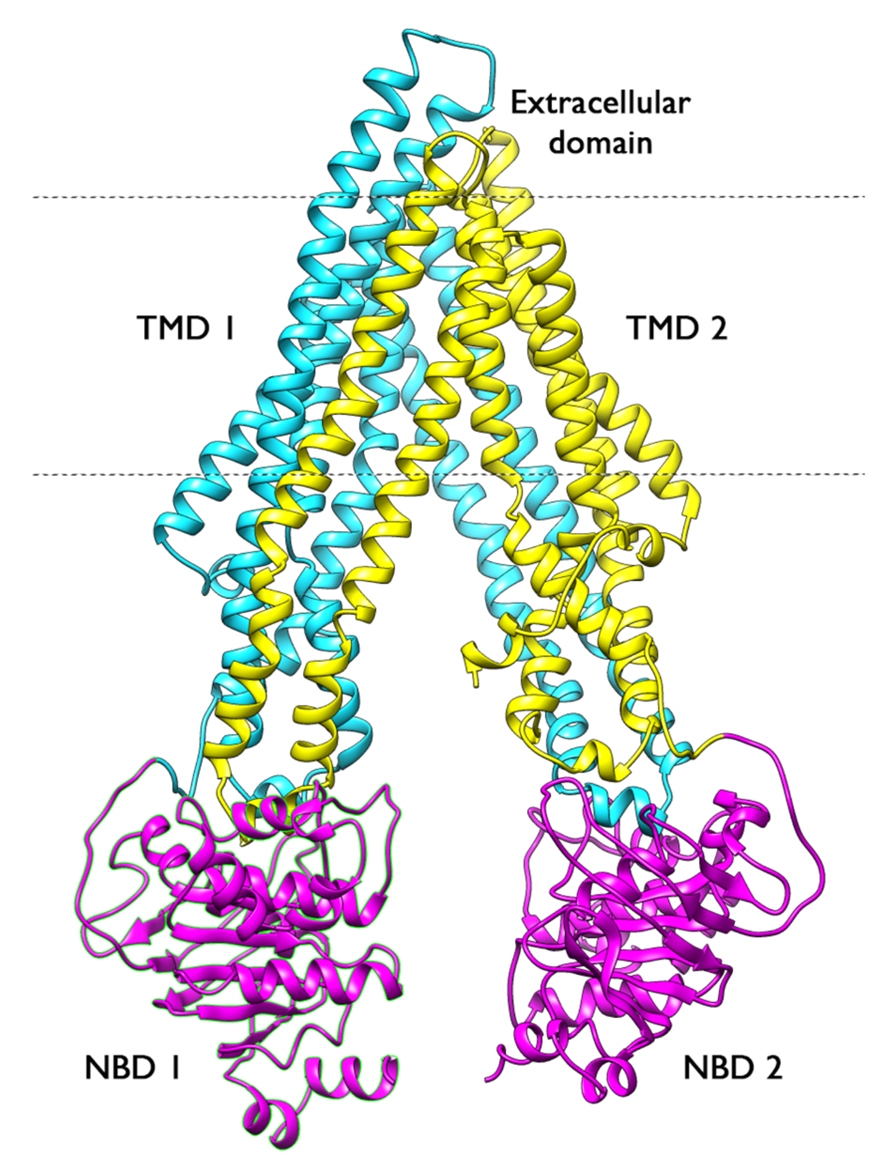
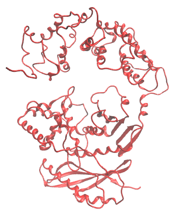
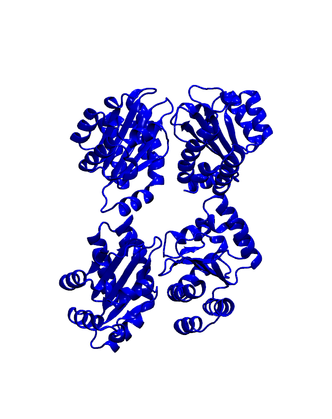
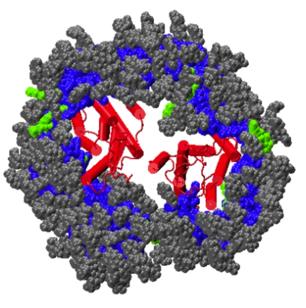
hepta-transmembrane domains (TMDs), the class C GPCRs specifically possess large amino-terminal extracellular domains (ECDs), which comprises of an 
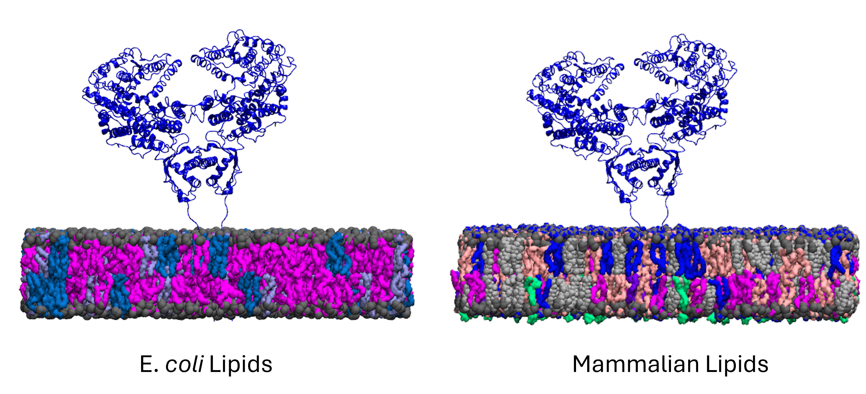
orthosteric binding region, a so-called Venus flytrap domain (VFT), and a cysteine-rich domain (CRD). The domains of mGluR1, specifically the 7TM domain has been shown to be regulated by its surrounding lipid environment, especially by cholesterol although the mechanism of regulation remains elusive.
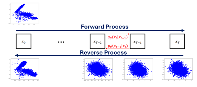
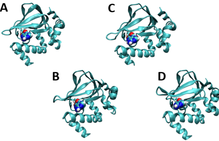
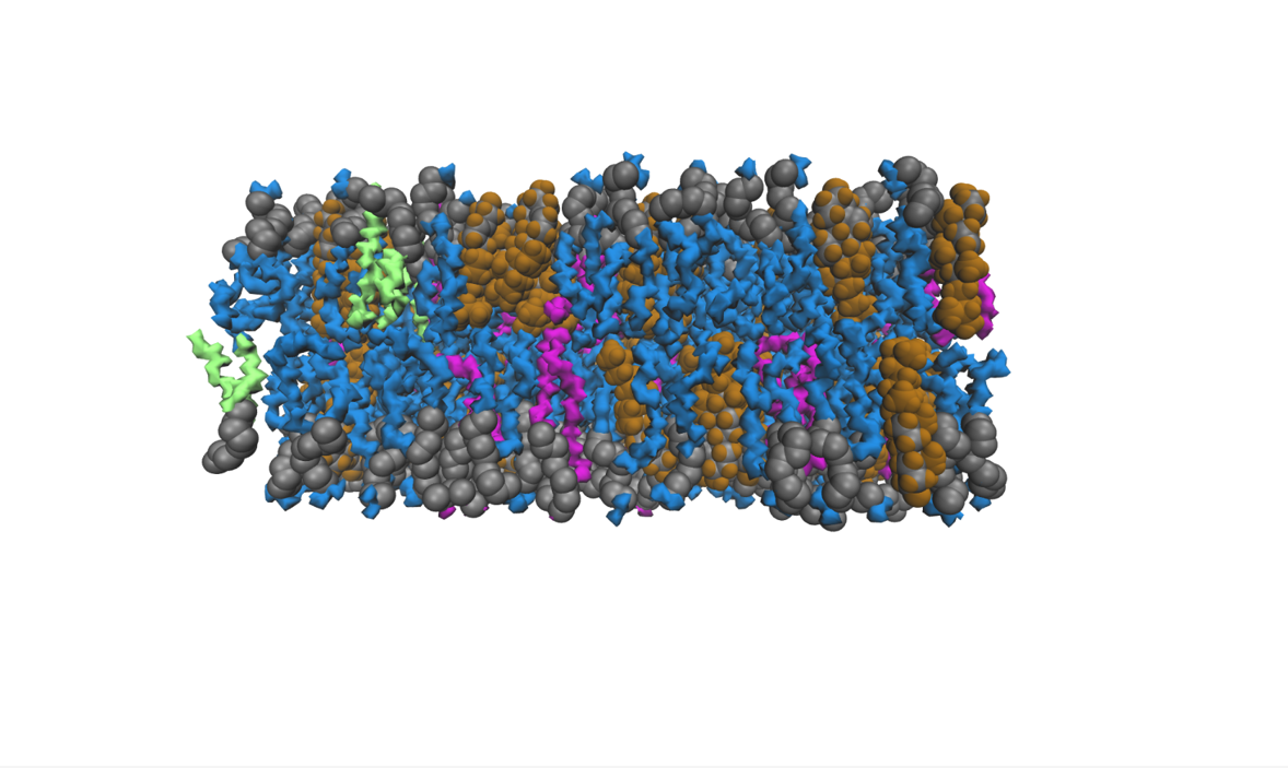
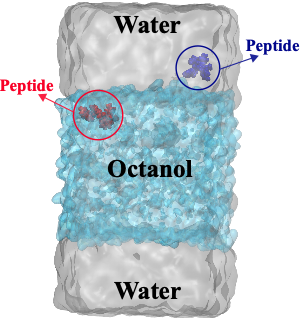
    To characterize the conformational dynamics of mGluR1, employing microsecond-level all-atom equilibrium and non-equilibrium MD simulations will enable us to determine the conformational rearrangements that occur in the different forms of this protein. Understanding and quantifying these functional changes could lead to the development of potent drugs targeting GPCRs. Due to high sequence conservation of the transmembrane domains (TMDs) of mGluRs, the molecular interactions observed showing cholesterol dependence in mGluR1 are likely to be applicable for other members of the mGluR family and possibly GPCR’s in general. Therefore, the goal of this research is to make novel, scientific, and significant contributions to drug development field by elucidating the activation mechanisms of G-Protein-coupled-receptors (GPCRs).
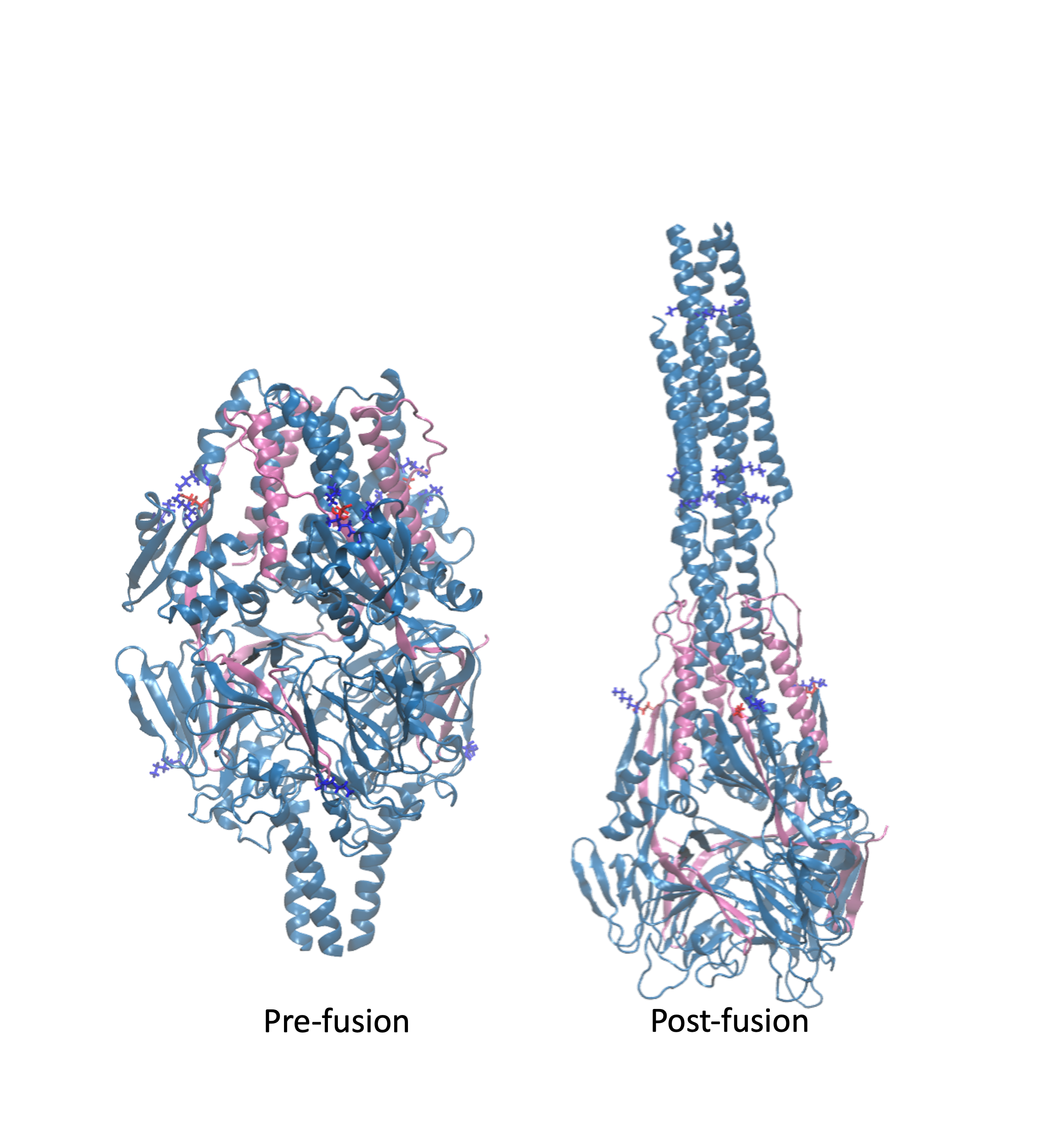
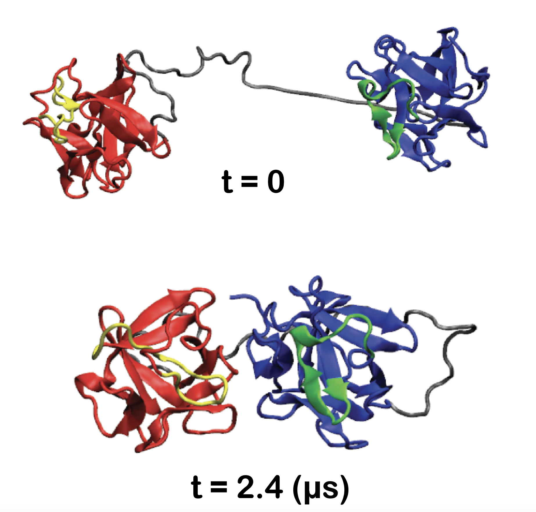
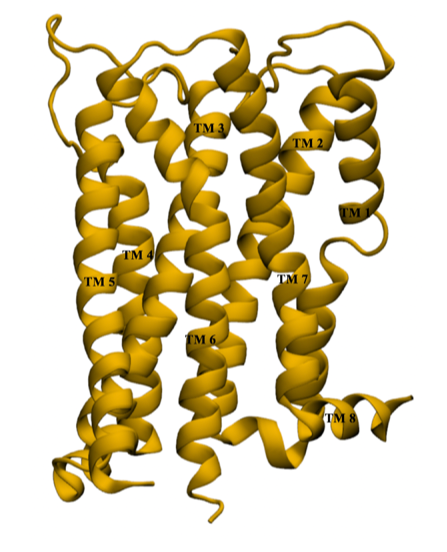
    Our findings have revealed that cholesterol influences the conformational changes of the internal protein and acts less significantly on individual protomers. We have also observed that cholesterol affects the dynamics of mGluR1 essentially in TM1 and TM2 which make up the inter-phase of the protein. 
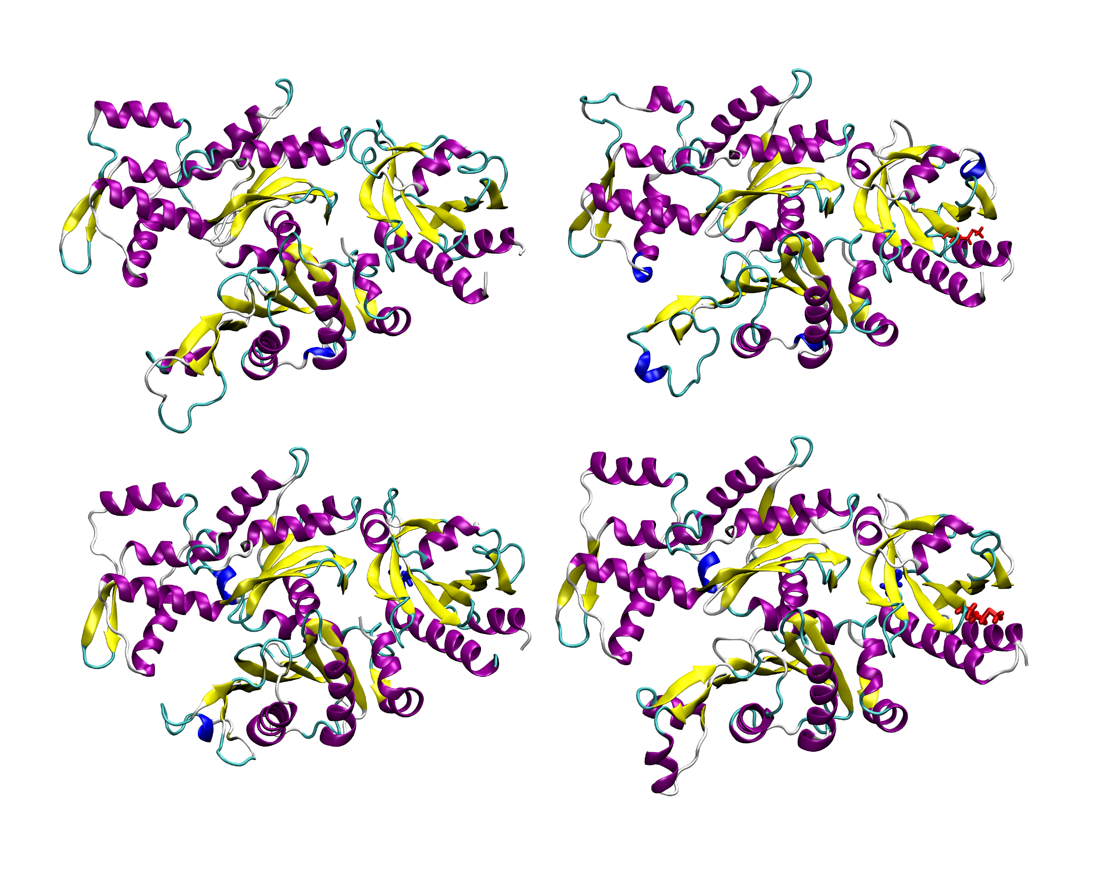
Further analysis shows that a low cholesterol concentration induces more significant conformational changes in mGluR1, while the system with higher cholesterol 
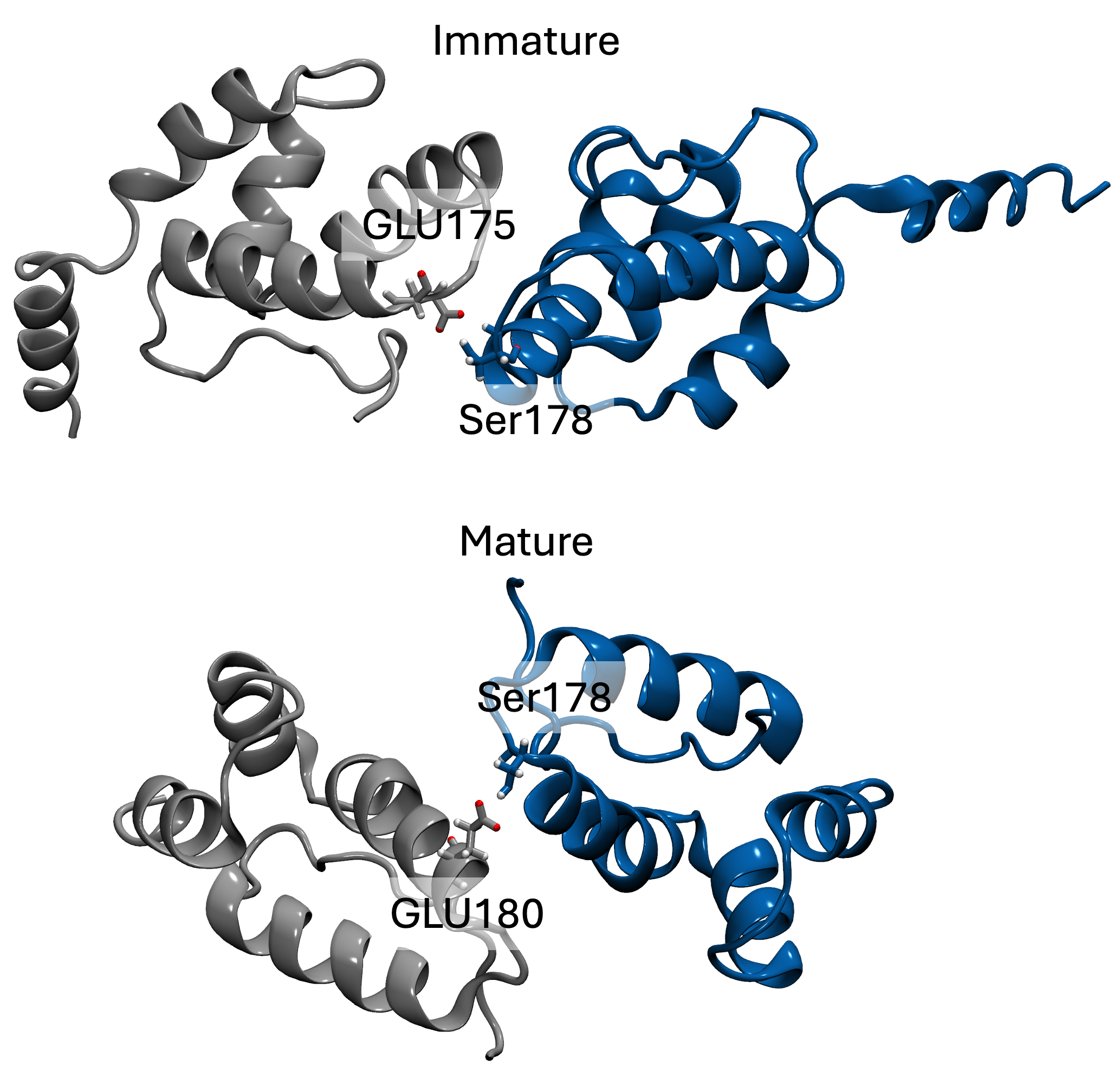
tends to behave similarly to systems without cholesterol. We have also identified some significant electrostatic interactions which are formed only in lower cholesterol concentration but absent in the other systems.

    Conference Poster:
    Cholesterol Dependence on the Conformational Changes of Metabotropic Glutamate Receptor 1 (mGluR1)

    Utility of a Time-lagged Autoencoder for Calculating Free Energy by Generating a Large Number of Synthetic Trajectories Based on Molecular Dynamics

    Using a Time-lagged Autoencoder for Calculating Free Energy Using MD Trajectories


    In this project, we present the time-lagged autoencoder (TAE) machine learning algorithm to predict a two-dimensional molecular dynamics trajectory from a Langevin Dynamic model. Regular molecular dynamics is very time-consuming since it needs to solve the Newtonian equation for millions of atoms in each time step. In TAE, the dimensionality of the problem is reduced and as a result, it can save time. This proposed novel use of the TAE algorithm provides a straightforward method of using deep learning in amplifying rare events and estimating accurate free energies in biomolecular systems.

    Conformational Dynamics of SARS-CoV-2 and its Variants of Concern

    Atomic-Level Characterization of SARS-CoV-2 and its Variants of Concern Using Molecular Dynamic Simulations

    Over three years since late December 2019, a rapidly expanding and evolving pandemic has claimed lives of millions and crippled economies around the world. SARS-COV-2, the virus behind the pandemic, uses its spike proteins to infect human cells by binding to ACE2 receptors on host cells. Due to the sheer volume of global infections, multiple mutations have arisen, among which there have been successful variants with increased transmissibility and capability for immune system evasions.
    Static information on the structure of wild-type spike proteins is not sufficient to understand the evolving process of infection by the coronavirus. Here we have used microsecond-level molecular dynamics (MD) simulations to study the important conformational dynamics of active and inactive states of the spike proteins from the wild-type, Alpha, Beta, Gamma, Epsilon, Delta, and Omicron variants of SARS-CoV-2, as well as an engineered spike protein associated with the Moderna vaccine.
    We have identified important mutations that contribute to changes in the structural and conformational dynamics of the spike protein. Our simulations reveal that certain mutations create significant instability in the spike protein. Notably, mutations shared by the Delta, Epsilon, and Beta variants appear to be responsible for the differential dynamic behavior observed in our simulations, which in turn could be linked to higher transmissibility and potential for immune evasion. This study provides insight into the dynamic behavior of the spike protein from different SARS-CoV-2 variants and could be used to identify targets for the development of novel vaccines and therapeutic agents.

    Characterization of the Conformational Dynamics of the RSV Fusion Glycoprotein

    Investigation of the RSV Fusion Protein Using Molecular Dynamics

    Respiratory syncytial virus (RSV) is a leading cause of acute lower respiratory infection in infants, young children, and older adults. The RSV fusion (F) glycoprotein is responsible for mediating the fusion between the viral envelope and host cell membrane, a process vital to viral infection. This process requires RSV F to undergo a significant conformational transition from the pre-fusion to the post-fusion conformational state. RSV F is the target of most known neutralizing antibodies, underscoring its dual significance mechanistically and therapeutically. Understanding the structural dynamics of RSV F is important for understanding the mechanism of viral infection and for drug and vaccine development for RSV infection.
    The primary objective of this research is to elucidate the conformational dynamics governing the transition of RSV F from its pre- to post-fusion states, offering atomic and thermodynamic insights. Unlike other class I fusion proteins activated by low pH, receptor binding, or interaction with a secondary viral glycoprotein, evidence suggests that RSV F is not activated by any of these events. It is possible that membrane fusion for RSV does not rely on a specific trigger but rather on the spontaneous conversion of RSV F to its post-fusion form. However, the energetics underlying these conformational changes and their relation to membrane fusion remain largely unexplored. The proposed research aims to fill this gap by providing detailed and reliable insights into the energetics of RSV F conformational changes.

    Investigation of P-glycoprotein Transport Cycle Using Molecular Dynamics

    Investigation of P-glycoprotein Transport Cycle Using Molecular Dynamics

    Drug resistance to chemotherapeutic drugs is a very challenging problem that often results in clinical failure of highly effective medications. A major mechanism of drug resistance can be attributed to the multi-drug resistance transporters that efflux drugs from the cytosol to the extracellular space. P-glycoprotein is a transmembrane ABC transporter that plays a crucial role in the reduction of intracellular concentration of a diverse range of xenobiotics that can vary tremendously in net charge, molecular size, and physicochemical properties.
    This study aims to utilize molecular dynamics simulations to understand the local and global conformational changes of the inward and outward states of P-glycoprotein.

    Comparison Study of Hyperpolarization-activated Cyclic Nucleotide-gated Channel Isoforms 1-4

    Atomic-Level Characterization of the Binding Activities of Hyperpolarization-activated Cyclic Nucleotide-gated Channel Isoforms Using Molecular Dynamic Simulations

    Hyperpolarization-activated cyclic nucleotide-gated (HCN) channels are a subfamily of tetrameric, voltage-gated, cyclic-nucleotide modulated K+/Na+ channels expressed in the heart and central nervous system. Referred to as “pacemaker” channels, they function as modulators of spontaneous rhythmic electrical activity. The binding of cAMP to the cyclic nucleotide binding domains (CNBDs) is able to induce a slight depolarizing shift in the voltage threshold necessary for channel activation, but on its own is not sufficient to open the channel in the absence of a hyperpolarizing membrane potential.
    Using atomistic molecular dynamics, we have simulated the isolated CNBD of HCN1-4 bound to cAMP and have used both equilibrium simulations and alchemical free energy perturbation to characterize and compare the binding activities of the different isoforms as well as the individual contributions made by specific residues. We hope to establish a molecular basis for the experimentally demonstrated difference in sensitivities and selectivities for cyclic nucleotides that exist within the subfamily.

    Molecular Dynamics Study of the FGF1/FGF2 Heterodimer in Human Fibroblasts

    Molecular Dynamics Study of the FGF1/FGF2 Heterodimer in Human Fibroblasts

    Fibroblast growth factors (FGFs) are a family of mitogenic proteins that regulate cell division, growth, and tissue repair. FGFs are classified into seven subfamilies based on phylogeny and sequence homology. The FGF1 subfamily (FGF1 and FGF2) is extensively studied and plays a crucial role in cell proliferation and angiogenic activity. However, FGF1 and FGF2 have low thermal and proteolytic stability, resulting in a short half-life under physiological conditions. To overcome these limitations, we modeled a fused protein combining FGF1 and FGF2 and conducted molecular dynamics simulations to study several engineered protein variants. Our observations revealed that each monomer within the FGF1-FGF2 dimer protein exhibited increased stability, regardless of heparin exposure. This enhanced stability resulted in improved biological functions and achieved high thermal stability. Furthermore, heparin-induced conformational changes facilitated the interaction of FGF1 and FGF2 with receptors, initiating signaling pathways.

    Human Immunodeficiency Virus Type 1 Envelope Glycoproteins

    Atomic-Level Characterization of the Human Immunodeficiency Virus Type 1 Envelope Glycoproteins Using Molecular Dynamic Simulations

    The human immunodeficiency virus (HIV) attacks the body’s immune system and if left untreated can progress into acquired immunodeficiency syndrome (AIDS) which leads to the inability to fight infection. The most common form of the virus, human immunodeficiency virus type 1 (HIV-1), enters host cells after the envelope (Env) glycoprotein trimer [(gp120/gp41)]3, a class I fusion protein, binds the CD4 receptor and a coreceptor, either CCR5 or CXCR4. Binding to the CD4 receptor induces conformational changes that allow mature (cleaved) Env to transition from a closed pretriggered state (state-1) to open states that are needed for the fusion process (state-2 and state-3). However, the closed conformational state of the Env trimer is not yet known.
    This study aims to characterize local and global conformational changes of the closed and open states of the Env trimer and to determine the dynamic behavior of the glycoprotein trimer through a combination of equilibrium and nonequilibrium molecular dynamic (MD) simulations. Additionally, the long-term goal of this project is to determine the pretriggered closed state (state-1) of the HIV-1 Env protein through the use of steered molecular dynamics (SMD).

    Cholesterol Concentration Effects on Bilayer Membranes and its Role in Designing Efficient Liposomal Drug Delivery Systems

    Studying the Effects of Varying Cholesterol Concentrations on Bilayer Membranes

    Liposomal drug delivery systems provide a versatile therapeutic platform for treating numerous diseases. Cholesterol, as a common component of many liposomal drug delivery systems, plays a crucial role in enhancing mechanical strength and decreasing the permeability of biomembranes.
    In this study, all-atom molecular dynamics simulations are performed to characterize the effect of cholesterol concentration in both planar and spherical bilayers. Furthermore, it has been quantified how a range of bilayer properties, including area per lipid, leaflet interdigitation, membrane thickness, and lipid Scd order parameters, are altered by variation in concentrations of lipid and cholesterol. Therefore, the most suitable amount of cholesterol and lipid to prepare stable and controlled drug release vehicles is obtained by screening the lipid and cholesterol ratio arrangement. Compared to membranes without cholesterol, the influence of cholesterol on lipid bilayers is revealed. These results provide a better understanding of the fundamental characteristics of the structure and dynamics of cholesterol-containing membranes. By revealing molecular details of interactions between cholesterol and phospholipids, these simulations accurately represent atomic-level characteristics of the planar and spherical bilayers. The simulations presented here shed light on the workings and limitations of liposomal drug delivery technology and establish a computational framework for the rational design of liposomes in drug delivery systems.

    Characterizing the Conformational Dynamics of SARS-CoV-2 Spike Protein Using smFRET

    Integration of smFRET and MD to Characterize Conformational Dynamics of the Spike Protein of Wild-type SARS-CoV-2 and its Variants

    Since the rise of the COVID-19 pandemic, researchers have pushed for development of efficient therapeutics and vaccines to assist in preventing the severity of its viral effects. A major target has been the trimeric SARS-CoV-2 spike protein. There has been various experimental and computational efforts to characterise the structural dynamics of this protein at different stages of its mechanisms. Among these methods are single molecule fluorescence resonance energy transfer (smFRET) experiments as well as molecular dynamics (MD) simulations.
    The purpose of our current work here is to combine the existing data from smFRET experiments and MD simulations to provide a more complete picture of prefusion SARS-CoV-2 spike protein conformational dynamics. We have particularly examined the conformational dynamics of several variants of this protein in a comparative manner. Specifically, we have examined multiple computational methods based on “implicit screening” approach within different approximations to predict dye-dye distance and FRET efficiency distributions to provide reliable interpretations for both smFRET experimental data and MD simulation data of various spike protein variants including the parental-type, the D614G mutation in the wild-type, the alpha, beta, gamma, delta, and omega variants as well as the engineered spike protein used in the Moderna vaccine.
    This work has yet to be concluded, but the hope is that results will shed light on the structural heterogeneity of the spike protein in different variants of SARS-CoV-2.
    Conference Poster:
    Analysis of the SARS-COV-2 Spike Protein Tagged With Dye, CY3 and CY5, Attachment Under smFRET Environment

    Lipid-Dependent Alternating Access Mechansim of Sav1866

    Atomic-Level Characterization of Sav1866's Alternating Access Mechansim Using Molecular Dynamic Simulations

    This study investigates the influence of the surrounding environment on the conformational changes of active membrane transporters. These transporters undergo conformational alterations between an inward-facing (IF) and an outward-facing (OF) state in order to facilitate the movement of substrates across cellular membranes. Our research focuses on a bacterial multidrug ABC exporter called Sav1866, and we employed microsecond-level all-atom MD simulations to explore the lipid-dependent nature of the alternating access mechanism.

    Rational Design Approaches to Liposomal and Lipid Nanoparticle-based Drug Delivery Systems

    By providing a drug release mechanism, channel-functionalized liposomes can offer a promising remedy to the drug delivery problem, particularly in delivering drugs into cancer cells. Liposomal and lipid nanoparticle-based therapeutics have been used to treat diverse diseases often offering reduced toxicity and enhanced efficacy compared to the active ingredients alone. Liposomes and lipid nanoparticles can in principle be engineered to release the drugs in response to certain stimuli such as acidity which is found in the tumor microenvironment. Although drug delivery liposomes and lipid nanoparticles are already being used in commercial medicines, targeted drug delivery has been significantly limited mainly due to the lack of an efficient method of triggering the drug release in response to a target-specific stimulus change. Despite much progress, the lack of a detailed, molecular-level picture of the complex processes involved in drug delivery liposomes and lipid nanoparticles' circulation in the bloodstream and their drug release mechanism has hindered the design of efficient drug delivery mechanisms.
    The overall objective here is to design robust simulation protocols to accurately describe lipid-nanoparticle/liposomal drug delivery at an atomic level to decipher the molecular basis of drug release. We also propose to use state-of-the-art all-atom molecular dynamics simulations in conjunction with statistical mechanics-based enhanced sampling techniques to provide a detailed, reliable activation process in a realistic liposomal and lipid nanoparticle environment. The rationale for the proposed research is that if the molecular basis of liposomal and lipid nanoparticle-based targeted drug delivery is better understood, drug delivery systems can be engineered more efficiently. It is anticipated that completing the study will demonstrate the importance of rational computational approaches, especially those providing multiscale structural and functional information of liposomes and lipid nanoparticles, to design novel and powerful liposome and lipid nanoparticle-based delivery systems.

    Conformational Dynamics of Cannabinoid Receptor 1 (CB1) in Active and Inactive Models

    Characterizing the Role of Chemo-mechanical Couplings in the Activation Process of CB1 Receptors Using MD Simulation


    Cannabinoid receptor (CB1), a member of class A G protein-coupled receptors (GCPRs), is a key component of the endocannabinoid system found in brain tissues. The CB1 receptor has been crystallized in active and inactive states and most recently, a Cryo-Electron Microscopy (Cryo-EM) structure has also been determined. Through the use of these structures, there has been some advancement in understanding cannabinoid receptor activation. However, a detailed knowledge of the dynamics involved in the activation mechanism remains elusive. Using equilibrium all-atom molecular dynamics (MD) simulations and using all three determined structures of CB1 receptor, we study the structural dynamics of the 7TM region of the CB1 in both x-ray crystal structures and Cryo-Em structures.
    The conformational transition of the protein from its inactive to active state is essential to understand the functionality of CB1 receptor. All-atom MD has been implemented to study the protein’s stability and conformational changes in both X-ray and Cryo-Em structures of CB1 receptor from its inactive state to the active state. The goal of this research is to characterize the role of chemo-mechanical couplings in the activation process of CB1 receptors. This study will shed some light on the conformational distinctions of the X-ray and Cryo-EM structures of CB1 receptor and provide a framework for comparing the activation process of CB1 receptor as compared to other GPCRs from a structural point of view.
    Our initial findings show that the Cryo-EM CB1 structure is a highly flexible system as opposed to the X-ray crystal structures. This is an on-going research and further findings will be updated.

    Conference Poster:
    Comparing the Dynamic Differences between X-ray and Cryo-EM Structures of CB1 Receptors using Molecular Dynamics Simulation

    Molecular Dynamics Simulation of the Effects and Mechanisms of Lipid Nanoparticles in Drug Delivery Systems

    Studying the Effects and Mechanisms of Lipid Nanoparticles in Drug Delivery Systems Using Molecular Dynamics


    Lipid nanoparticles (LNPs) have emerged as promising carriers for delivering various therapeutic molecules, including nucleic acids and small molecules because they have the ability to protect encapsulated drugs from degradation and clearance by the body’s immune system. Another crucial aspect of LNPs is their bio-compatibility as they do not have any adverse effects on human cells. They are important to work with because they allow us to easily modify their composition to alter their capabilities of binding.

    Dynamical Effects of ALS-associated Mutations on Profilin

    MD Simulation Study of Mutations in Profilin and their Effects on ALS


    Amyotrophic Lateral Sclerosis (ALS), known as Lou Gehrig's disease, has been on the rise to spproximately 31000 patients in the USA, according to CDC. It has been associated with specific mutations on Profilin, a protein with specific binding sites for both poly-L-proline (PLP) and Actin. To maintain high rates of actin filament growth cells keep an ample supply of actin monomers above the critical concentration needed for polymerization. Most of these monomers capable of polymerizing are in the form of complexes between Profilin and Actin. Profilin-1 (PFN1) (139 amino acids) is an actin-binding protein involved in actin filament elongation in cells. The X-ray crystal structure of PFN1 in a ternary complex with Actin (375 amino acids) and a poly-L-proline chain (13 amino acids) from VASP (vasodilator-stimulated phosphoprotein), was published in 2007 (Ferron et al., EMBOJ) - 2PAV – 1.8 angstroms. Mutations in PFN1 are implicated in amyotrophic lateral sclerosis (ALS), a neurodegenerative disorder. ALS-linked mutations destabilize Profilin. Mutant Profilin produces ubiquitinated insoluble aggregates.
    Using close to 30 microseconds of All-atom molecular dynamics simulations across 16 systems of study composed of Profilin, Actin, and PLP, we have found that individual Profilin variants do not show much difference in dynamics. However, the mutants slightly decrease the flexibility of the Profilin. The T109M mutation induces the largest change in the secondary structure of the Profilin. In complex with Actin, Profilin loses much more flexibility, specifically in the Actin-binding region. This may cause the aggregation of Profilin. The secondary structure of T109M is altered more significantly than other mutants. In the APV complex in the presence of PLP, both Actin, and Profilin lose flexibility in individual mutations (more in T109M) but double mutation. The double mutation gains similar flexibility with the WT. However, the full dynamics of Profilin is not resumed. We have identified intermolecular interactions that could be among the main reasons for conformational differences between wild-type Profilin and mutants and potentially the reasons for the ALS at the molecular level due to the mutations.

    Nanoparticle Synthesis

    Designing Recombinant Proteins for Peptide-Directed Nanoparticle Synthesis

    In this study, we investigate in a comparative manner how a specific peptide known as the Pd4 and its two known variants may form nanoparticles both in an isolated state and when attached to the green fluorescent protein (GFPuv). More importantly, we introduce a novel computational approach to predict the trend of the size and activity of the peptide-directed nanoparticles by estimating the binding affinity of the peptide to a single ion.

    I used molecular dynamics (MD) simulations to explore the differential behavior of the isolated and GFP-fused peptides and their mutants.
    Some questions I seek to address with this work:

    1) How Mutations in peptide effect the size and shape of the Nanoparticle?

    2) How GFP would control the peptide conformational dynamics?

    3) Can GFP overcome the mutational effect of peptide on Nanoparticle Size?

    4) Is secondary important for catalytic reaction activity?

    5) Estimating peptide amino acid and nanoparticle binding free energy helps understand Nanoparticle formation?
    The findings reveal that whereas isolated Pd4 and its two known variations (A6 and A11) form nanoparticles of various sizes, fusing these peptides to the GFPuv protein yields nanoparticles of identical size and activity. To put it another way, the GFPuv decreases the nanoparticles' peptide sequence sensitivity. This research develops a computational framework for creating free and protein-attached peptides, which aids in the creation of nanoparticles with well-controlled characteristics.

    Characterizing Mitochondrial Localization Peptide Isoforms Present on the Androgen Receptor Based on Secondary Structure Propensity

    Atomic-Level Characterization of Mitochondrial Localization Peptide Isoforms on the Androgen Receptor Using Molecular Dynamic Simulations

    The androgen receptor (AR) is a nuclear receptor that binds to androgenic hormones in the cytoplasm and is translocated into the nucleus where it functions as a regulator of DNA transcription. Recently, AR has been shown to be capable of both localization and import into the mitochondria as well, where it continues to play a regulatory role. This is possible due to the presence of a 36-residue mitochondrial localization sequence (MLS) at the N-terminus, which was demonstrated to be capable of targeting a passenger protein for mitochondrial import, and whose deletion was sufficient to prevent proper import of AR into the mitochondria. Analysis with MitoProt predicted the presence of an MLS and a cleavage site after the first 14 residues, and mutational studies are currently underway in this region.
    Using both biased and unbiased molecular dynamics simulations, we have simulated 4 experimental isoforms (E2A, E2K, E2Q, and wt) of the isolated, intrinsically disordered, 15-residue peptide, dubbed the mitochondrial localization peptide (MLP). We are developing a scheme for characterizing IDPs based on their secondary structure propensity, which we will use to understand how these mutations affect the structural/conformational dynamics of the peptide. We hope to use these findings to explain the observations of our collaborators, as well as to suggest potential mutations of experimental interest that will inform our understanding of the mechanisms of protein localization/import to the mitochondria.

    Mechanosensitive Channel of Large Conductance (MscL)
    Spontaneous Activation

    Applying Biased and Unbiased Molecular Dynamics

    Understanding the chemical basis of an engineered mechanosensitive channel's spontaneous activation is important for engineering more efficient pH-sensitive mechanosensitive channels. The work provides a computational framework to accomplaish this goal by using an orientation-based strategy used to generate and optimize an open model of modified MscL. This is a promising tool for creating unknown protein states and exploring ion channel activation mechanisms. This study aids research into pH-triggered drug delivery liposomes (DDL), which use MscL as a nanovalve.

    Research article:
    Elucidating the molecular basis of spontaneous activation in an engineered mechanosensitive channel

    An Investigation of the YidC-Mediated Membrane Insertion of Pf3 Coat Protein

    Mechanistic Study of Protein Mechanism Pathway


    YidC is a membrane protein that facilitates the insertion of newly synthesized proteins into lipid membranes. Through YidC, proteins are inserted into the lipid bilayer via the SecYEG-dependent complex. Additionally, YidC functions as a chaperone in protein folding processes. Several studies have provided evidence of its independent insertion mechanism.
    The mechanistic details of the YidC independent protein insertion mechanism remain elusive at the molecular level. In this study, we looked at the local and global conformational changes of YidC associated with Pf3 insertion into the hydrophilic groove, Pf3 interactions with YidC and the membrane, and conformational changes in Pf3 that occurred during the insertion process.
    The incoming Pf3 coat protein will first get intact with the hydrophilic groove located in the trans-membrane region forming a salt-bridge with ARG72. The positive charged arginine will form a salt bridge with the negative charged amino acid of the Pf3 coat protein D7. This salt bridge formation play a very big key role in the insertion mechanism of YidC. The Pf3 coat protein then moves towards the periplasmic side of the membrane due to the force of a hydrogen bond attraction with the E2 region of the YidC protein. This interaction will help guide the protein movement towards the periplasmic side which is also assisted by the salt-bridge interaction between D18 of Pf3 and ARG72 of YidC. The combination of these interactions will stabilize the Pf3 protein position in the membrane. On other hand, for pushing the Pf3 protein in to the hydrophilic groove of the YidC, the loops of YidC on the cytoplasmic side of the membrane are very crucial. Since there are interactions that occur within this region, we believe that the cytoplasmic loops form a strong contact with the Pf3 coat. Overall, the protein is able infiltrate across the membrane within water-filled cleft, leaving the adjoining hydrophobic TM region in the lipid bilayer.

    Conference Poster:
    Membrane Insertion Of A Pf3 Coat Protein Using MD Simulations

    Atomic Level Characterization for the Transport Cycle Pathways of Bovine Multidrug Associated Protein 1 (bMRP1)

    Molecular Dynamics Characterization for the Transport Cycle Pathways of Bovine Multidrug Associated Protein 1 (bMRP1)

    Bovine Multidrug Resistance-Associated Protein 1 (bMRP1), belonging to the ABC family of membrane transporters, plays a crucial role in clinical settings. It is highly expressed in cancer cells, and its overexpression is closely associated with resistance to a broad spectrum of chemotherapeutic agents, often resulting in treatment failure and disease progression among cancer patients. This resistance phenomenon extends to leukemia, breast, lung, ovarian, and other solid tumors, where MRP1 utilizes ATP energy to actively pump out anticancer drugs from within cancer cells, significantly diminishing their therapeutic efficacy. Consequently, understanding the intricate role of MRP1 in drug resistance has become paramount in the ongoing quest to develop novel anticancer drugs, with researchers passionately exploring strategies to bypass its impact on chemotherapy resistance.
    The central goal of this research is to unravel the most probable pathway of the transitions between conformational states for bMRP1's full transport cycle with substrate included. Furthermore, we will characterize the interplay between different lipids in the cell membrane and MRP1.

    Developing Efficient Transfer Free Energy Calculation Methods
    For Hydrophobicity Predictions

    Molecular Dynamics for Hydrophobicity Prediction

    The interaction of peptides with membrane lipids is significant in biological processes. Short peptides are an excellent alternative to immune response antibodies, and they play a very crucial role in binding, insertion, and folding of membrane proteins. The characterization of solvent dependent conformational ensemble of the peptides is required for a molecular-level understanding of the thermodynamic hydrophobicity scale. To characterize the solvent-peptide interactions, we have developed a computational procedure that allows us to accurately model the peptides in both aqueous and organic solvent conditions and determine their properties at a thermodynamic level. This study evaluates the peptide conformational dynamics at different temperatures using molecular dynamics (MD) in the explicit solvent of water and octanol to estimate the transfer free energies accurately and to predict the partition coefficients. We have used a series of equilibrium MD simulations, and alchemical free energy calculations to measure the transfer free energies within various approximations. This study sheds light on the efficiency and accuracy of several different computational strategies for the study of transfer free energies.

    Conference Poster:
    Developing Efficient Transfer Free Energy Calculation Methods For Hydrophobicity Predictions

    Molecular Dynamics Investigation of the Influenza Hemagglutinin Conformational Changes

    Molecular Dynamics Simulationn of Influenza Hemagglutinin

    Hemagglutinin (HA) is a homotrimeric glycoprotein located on the surface of influenza virus and mediates fusion between viral and endosomal membranes of the host cell. The HA is synthesized as inactive HA0. HA0 is cleaved into HA1 and HA2 glycopolypeptides by host proteases. When the pH within the endosome drops, the HA2 protein undergoes a large irreversible conformational change in HA, while HA1 moves away from the HA2 domains, allowing the refolding of the HA2 loop into a helix and releasing the fusion peptide at the HA2.
    In this study we are investigating the conformational changes of HA2 trimer in the absence of HA1 under various pH condition through protonation of zero, one, two, or three conserved Histidine residues located on hinge region of HA2 using all-atom molecular dynamics simulations. This study aims to simulate HA2 with different protonation states.
    According to our current trajectory for these eight models, it appears that HA2 is undergoing conformational changes such as breaking and forming Hydrogen bonds and salt bridges specially between HSP and negative residues even when a single histidine residue is protonated. However, longer simulations may indicate more differential dynamic behaviors between different protonation states.

    Conference Poster:
    Molecular Dynamics Investigation of the pH-dependent Influenza Hemagglutinin Conformational Changes

    Investigation of PCAT and MsbA in the Presence and Absence of Magnesium

    Comparison of the Relative Free Binding Energies of PCAT and MsbA in the Presence and Absence of Magnesium Using Molecular Dynamic Simulations

    Peptidase-containing ATP-binding cassette (ABC) transporters are a family of bacterial ABC transporters that include a peptidase domain (PD). ABC transporters follow an alternating access mechanism to transport their native cargo, where the binding of ATP induces a conformational shift from an inward-facing (IF) state to an outward-facing (OF) state, and the subsequent hydrolysis and release of ATP reverts the protein to its inward-facing state. Recently, collaborators at Vanderbilt University have experimentally observed several unusual behaviors in PCAT1 that distinguish it from other ABC transporters, including differential magnesium titration behavior, and the ability to sustain reduced hydrolytic activity of ATP even after the mutation of a highly-conserved residue known to be necessary for catalysis in similar transporters (E648Q). Conclusions are drawn relative to MsbA, which is used as a model ABC transporter.
    Using atomistic molecular dynamics, we have simulated both PCAT and MsbA bound to ATP in the presence and absence of magnesium. Using equilibrium simulations and alchemical free energy perturbation methods, we hope to compare the relative free binding energies of these channels for ATP/ADP in the presence/absence of magnesium and in different conformational states (IFc/IFo/OF), as well as the individual contributions made by specific residues. We hope to use these findings to establish a molecular basis for the differential behaviors for PCAT1 observed by our collaborators.

    Investigation of Quorum Sensing in Pseudomonas aeruginosa

    Characterization of Quorum Sensing Using Molecular Dynamic Simulations

    This project aims to investigate the molecular dynamics of quorum sensing in Pseudomonas aeruginosa, a bacterium known for its antibiotic resistance, by evaluating the mechanisms and properties of quorum sensing inhibitors through classical molecular dynamics. A primary focus of this project will be on analyzing disruption of the quorum sensing system—a communication mechanism bacteria use to regulate gene expression based on their population density. By understanding how these inhibitors interact with the signaling molecules of bacteria, the study aims to hinder their ability to coordinate virulence and biofilm formation, offering potential new pathways for treating infections caused by this pathogen, and potentially other pathogens as future work delves into new, novel ideas that could arise from the goal of this study.

    Influenza Hemagglutinin (HA) is a Paradigm for Protein-mediated Membrane Fusion

    Molecular Dynamics of Membrane Fusion

    The proposed research project aims to study the conformational dynamics of the complete trimeric influenza HA ectodomain for the first time at an atomic level. In particular, we will study the confor- mational changes of HA that are triggered by protonation of a highly-conserved histidine residue in the HA2 hinge region. This will be followed by an investigation of the conformational transitions of HA2 upon the full exposure to solvents due to removal of HA.

    Conference Poster:
    Molecular Dynamics Investigation of The Ph-Dependent Influenza Hemagglutinin Conformational Changes
    The long-term goal of this research is to develop a computational framework for the rational design of novel therapeutic agents targeting the conformational rearrangements that drive the influenza HA-mediated membrane fusion process. The overall objective of this study is to accurately describe, at an atomic and thermodynamic level, the structural dynamics of HA in neutral and acidic environments. By employing microsecond-level all-atom equilibrium and non-equilibrium MD simulations, the conformational rearrangements that occur when influenza HA is exposed to an acidic environment will be characterised.

    Conformational Free Energy Landscapes of
    SARS Coronavirus Spike Glycoproteins

    The State-of-the-art Enhanced Sampling Molecular Dynamics Simulations

    The proposed research project aims to study understanding how coronavirus spike glycoproteins undergo conformational changes to bind to host ACE2 receptors is key to the development of coronavirus vaccines and therapeutics, which requires a dynamic rather than a static picture to provide a reliable structure-based drug design framework. The virus that causes COVID-19, SARS-CoV-2, is more stable and slower changing than the previous form that caused the SARS outbreak in 2003, according to new computational simulations of the behavior of SARS-CoV-1 and SARS-CoV-2 spike proteins before fusion with human cell receptors.

    Conference Poster:
    Characterizing The Roles Of Chemomechanical Couplings In The Differential Behavior Of Sars-cov-1 And Sars-cov-2 Spike Glycoproteins

    Stability of mGluR2 in Micelles: Role of Branched Detergents

    Using Molecular Dynamics to Study mGluR2 Stability in Micelles

    Metabotropic glutamate receptors (mGluRs) are transmembrane proteins composed of seven α-helices and belong to the G-protein coupled receptor (GPCR) family. With over 800 identified entries, the GPCR family is one of the largest gene families in the human genome. Among these entries, eight distinct members (mGluR1–8) have been identified, exhibiting variations in their affinity to glutamate and their mode of G-protein coupling. Within this group, the metabotropic glutamate receptor 2 (mGluR2) plays a crucial role in the central nervous system. Interestingly, mGluR2 has been observed to be influenced by its neighboring lipid environment, particularly cholesterol, although the exact mechanism behind this regulation remains unknown. Moreover, micelles are widely utilized in research to enhance solubility and stabilize hydrophobic molecules, including membrane proteins like mGluR2. Incorporating mGluR2 into a micelle offers several advantages in biochemical and biophysical studies.

    This study aims to enhance solubility, characterize the structure, conduct functional analysis, and explore drug discovery applications through the incorporation of mGluR2 into a micelle. These objectives collectively contribute to a comprehensive understanding of mGluR2 and its potential implications in various biological processes and therapeutic development.

    The Effect of Cholesterol Concentration on Lipid Bilayers: A Coarse-grained Molecular Dynamics Study

    Using Molecular Dynamics to Study the Effect of Cholesterol Concentration on Lipid Bilayers

    This study utilizes coarse-grained molecular dynamics simulations to investigate how cholesterol concentration influences planar and spherical lipid bilayers, essential in liposomal drug delivery. Key findings highlight cholesterol's role in liposome shape and physical properties, offering insights into optimal cholesterol-liposome ratios for stable and controlled drug release. The research provides valuable atomic-level details, advancing our understanding of drug delivery mechanisms.